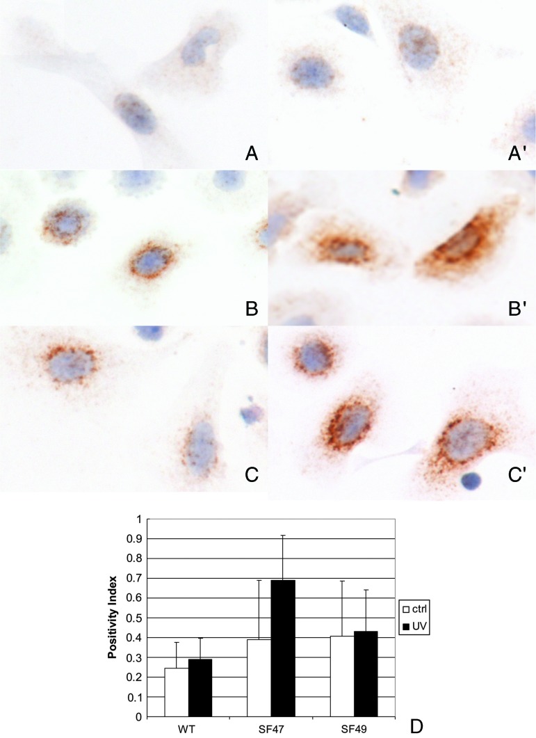
Figure 5.
Furin expression in murine epidermal keratinocytes in primary culture detected with IHC. Panels A, B, and C correspond to unirradiated cultures derived from WT SKH-1, SF47, and SF49 mice, respectively. Similarly A′, B′, and C′ correspond to UVB-irradiated WT (SKH-1)-, SF47-, and SF49-derived cultured keratinocytes. The cells were irradiated with 0.5 kJ as described in the text and evaluated 15 minutes post-exposure. Furin IHC and hematoxylin counterstain x 1000. Panel D shows image analysis values of furin positivity in the cytoplasm using a quantification procedure based on computerized image analysis as described in the Materials and Methods section.

