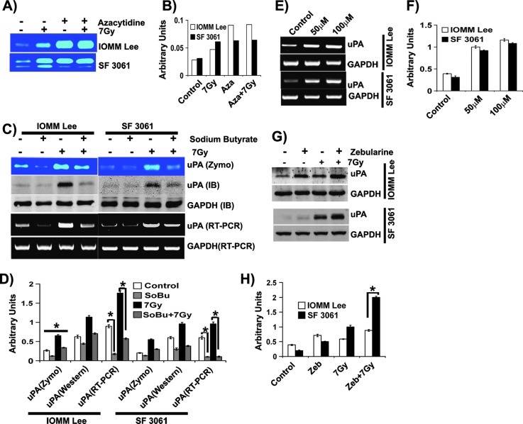
Figure 2.
Hypomethylation is linked to uPA expression in meningioma. (A) IOMM-Lee and SF3061 (1 x 10⁵) cells were treated with azacytidine (10 µM) for 24 hours before irradiation followed by incubation in serum-free medium overnight. Conditioned media were collected and 0.1 µg of protein was analyzed for uPA activity using fibrin zymography. (B) The band intensities were quantified and represented as arbitrary units. (C) IOMM-Lee and SF3061 (1 x 10⁵) cells were treated with sodium butyrate (2 mM) for 24 hours before irradiation. Conditioned media were subjected to fibrin zymography, total RNA was used to perform RT-PCR, and cell lysates were used for immunoblot analysis. (D) The band intensities were quantified and represented as arbitrary units. (E) IOMM-Lee and SF3061 (1 x 10⁵) cells were treated with zebularine for 24 hours. Total RNA was used to perform RT-PCR, and cell lysates were used for immunoblot analysis for uPA. (F) The band intensities were quantified and represented as arbitrary units. (G) Meningioma cells were pretreated with zebularine (50 µM) for 24 hours, and the cell lysates were used to perform immunoblot analysis for uPA and GAPDH. (H) The band intensities were quantified and represented as arbitrary units. Each blot/image is representative of three independent experiments, and each column represents a mean of three values (asterisk represents significant value of P < .05).

