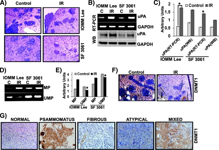
Figure 6.
Radiation treatment induces hypomethylation in vivo. Nude mice were intracranially implanted with IOMM-Lee cells as described in the Materials and Methods section. (A) Formalin-fixed brain sections were subjected to H&E staining and observed under x20 objective. Each image represents a brain section of five animals. (B) Total RNA was extracted from frozen brain tissues, and RT-PCR was performed for uPA and GAPDH. Tissue lysates were analyzed for uPA expression by Western blot analysis. (C) The band intensities of RT-PCR gels and Western blots were quantified and represented as arbitrary units. (D) Genomic DNA from frozen brain tissues was extracted, treated with bisulfite, and subjected to MSP targeting the uPA promoter (MP, methylated primer; UMP, unmethylated primer). (E) The band intensities were quantified and represented as arbitrary units. (F) Immunostaining with anti-DNMT1 antibody and non-specific IgG (NsIgG) was performed on formalin-fixed brain sections from animals implanted with IOMM-Lee cells followed by treatment with HRP-conjugated secondary antibody and DAB staining. Representative photomicrographs (x200) are shown in comparison to normal mouse brain sections. (G) Immunohistochemical analysis for DNMT1 was performed on tissue microarrays and clinical meningioma samples. Representative images (x400) are shown. Each column represents a mean value of brain tissues from three different animals (*P < .05, significant difference from respective normal tissues).

