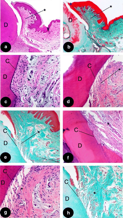
Fig. 4.

Details of different treatment groups. a Supra-crestal tissues (asterisk); showing random orientation of the fibers in the empty group (HE; original magnification, ×2.5), please observe the supra-crestal tissues are detached from the rootsurface, indicating a weak bond between the two tissues; b another example of an empty group sample (Masson; original magnification, ×2.5); c detail of a functionally orientated supra-crestal connective tissue attachment (asterisk), showing new cementum formation (c) in a EMD sample (HE; original magnification, ×20); d detail of a functionally orientated supra-crestal connective tissue attachment (asterisk), showing new cementum formation (c) in another EMD sample (HE; original magnification, ×10); e detail of a functionally orientated supra-crestal connective tissue attachment, showing new cementum formation in a sample treated with CaP/EMD (Masson; original magnification, ×10); f supra-crestal connective tissue attachment in CaP/EMD group (HE; original magnification, ×10); g higher magnification of (d) showing new cementum (c) (HE; original magnification, ×20); h higher magnification of (e) showing new cementum (c) (Masson; original magnification, ×20). C cementum, D dentin of the root. Asterisk, supra-alveolar tissues
