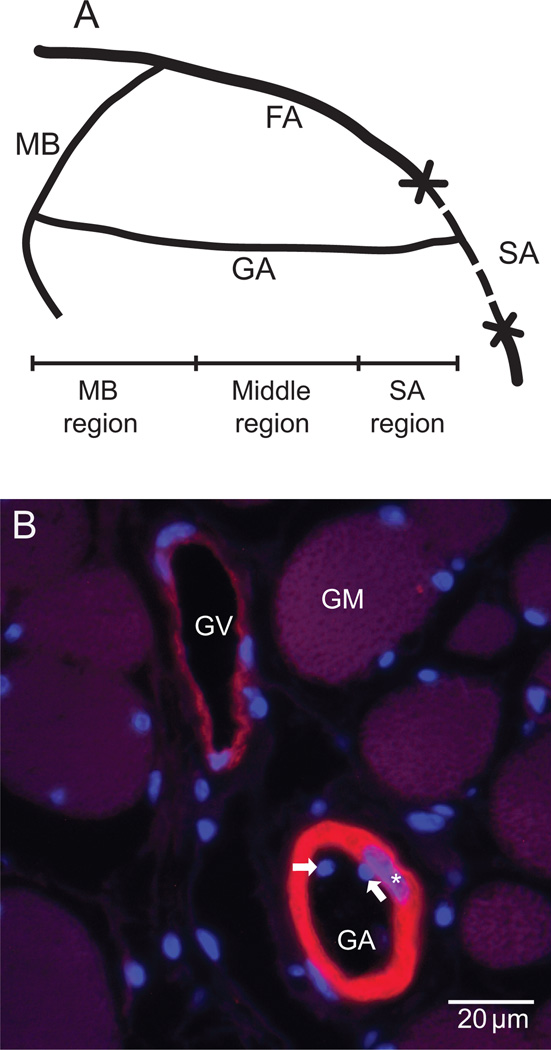
Figure 1.
A. Schematic of the surgical procedure and the three regions of the gracilis artery. FA: femoral artery. MB: muscular branch of FA. SA: saphenous artery. GA: gracilis artery. B. Micrograph of a paraffin section stained for smooth muscle and cell nuclei by immunohistochemistry using fluorescently labeled anti smooth muscle α-actin (red/yellow) antibody and bisbenzimidazole (BBI, blue), respectively. The medial cross-section area was estimated from measurements of the α-actin positive area. Endothelial cell nuclei are represented by the BBI positive structures inside the vessel lumen (arrows), while the smooth muscle cell nuclei are the BBI structures overlapping the α-actin positive areas (stars). GA: gracilis artery, GV: gracilis vein, GM: gracilis muscle fibers.

