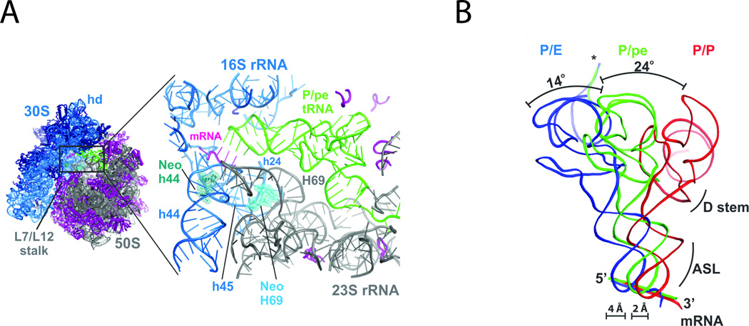
Figure 7.
The neomycin-bound ribosome. (A) Neomycin binds the small subunit (blue) in h44 (green) and the large subunit (grey) in H69 (light blue). (B) Ribosomes with neomycin bound at H69 display an intermediate position of P-site tRNA (green), between the hybrid (blue) and classical (red) configurations. Figure adapted from. (74)

