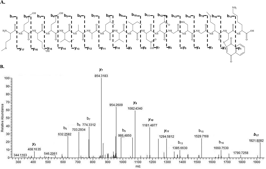
Figure 2.
Actin peptide MEEEIAALVIDNGSGMCK (1–18) adducted by 1,4-naphthoquinone with a MASCOT ion score of 60. A is the fragmentation pattern of depicted b- and y-ion series for the adducted peptide. B is the CID spectrum of [M+H]2+ at m/z 1033 (modified peptide); 1,4-NPQ is bound to the Cys17. Italicized ions y2, y7, and y9 – y11 are modified y-ions in the presence of the NPQ. Italicized b17 (m/z 1921.8082) represents the modified N-terminal fragment peptide. The peptide was doubly charged.

