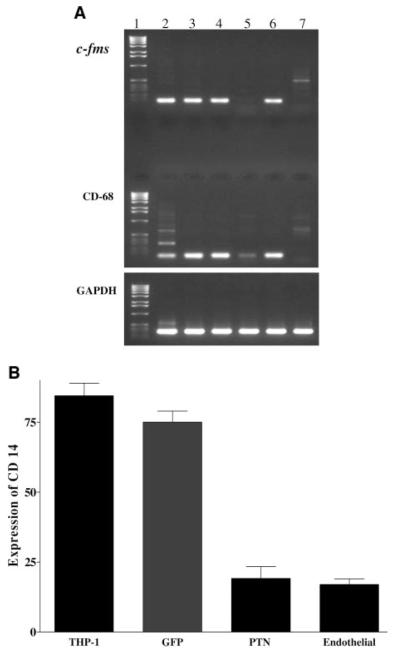
Figure 1.
PTN downregulates expression of monocytic cell markers. A, Total RNA was extracted from THP-1 cells grown in 10% serum (lane 2), induced to differentiate into macrophage-like cells by addition of 25 ng/mL phorbol 12-myristate 13-acetate (PMA; lane 3), transduced with retroviral bicistronic vector expressing: GFP (lane 4), PTN sense strand (lane 5), PTN antisense strand (lane 6) followed by treatment with PMA. The exponentially growing human coronary artery endothelial cells (lane 7) were used as a negative control. Analyzed monocytic cell markers were c-fms and CD-68 with primers predicted to amplify 97- and 132-bp DNA fragments, respectively. GAPDH primers were used as control a for the RT-PCR. Lane 1 is a DNA ladder marker. B, Flow cytometry analysis was performed by incubating 5×10⁵ THP-1 cells expressing PTN or GFP with phycoerythrin-labeled anti-CD14 antibody from PharMingen. Human coronary artery endothelial cells were used as a negative control. Uninfected THP-1 cells were used as positive control, and human coronary endothelial cells (endothelial) were used as a negative control. FACS analysis was performed at the Cedars-Sinai Research Institute Core Facility. Each experiment was repeated 3 times, and each bar graph represents mean±SEM of 3 experiments.

