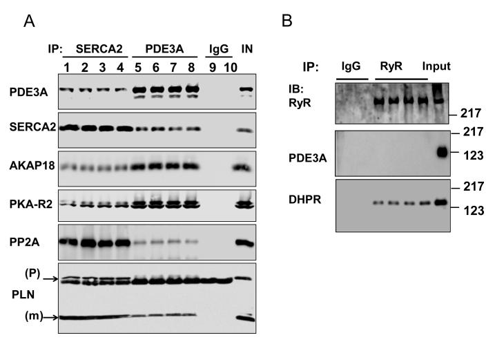
Figure 8. PDE3A co-immunoprecipitates with SERCA2 in murine cardiac tissue.
A) Representative Western blots from 2 way co-immunoprecipitation experiments, illustrating that PDE3A interacts with SERCA2a, PLN, PKA-RII and AKAP-18. As described in methods, precleared total WT heart lysates (1 mg) were incubated with anti-PDE3A (C terminal epitope) (3 μg) and anti-SERCA2a (3 μg) antibodies overnight at 4°C, followed by incubation with protein G beads (2h, 4°C). Immunoprecipitated proteins were eluted from the beads, and subjected to SDS PAGE/Western immunoblotting with the indicated antibodies. This experiment was representative of 3 independent experiments, (1heart/lane). B) Representative Western blots illustrating that PDE3A does not co-immunoprecipitate with the SR Ca2+ release channel (RyR2). Again, as described above, precleared total heart WT lysates (1mg) were incubated with anti-RyR antibodies, and immunuprecipitated proteins were subjected to SDS PAGE/Western immunoblotting with the indicated antibodies. This experiment was representative of 2 independent experiments, (1heart/lane).

