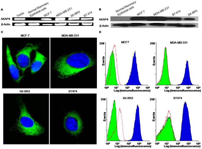
Figure 1. AKAP4 expression and localization in breast cancer cell lines.
A, Reverse-transcriptase PCR analysis showing AKAP4 gene expression in testis, all breast cancer cell lines, MCF7, MDA-MB 231, SK-BR3 and BT474 but not in normal human mammary epithelial cells. B, AKAP4 protein expression was detected by employing Western blotting. C, Cells were fixed, permeabilized and processed for indirect immunofluorescence studies which revealed cytoplasmic localization (green color) of AKAP4 protein. Nuclei were stained blue using DAPI. D, Flow cytometric analysis of live cells demonstrating surface expression of AKAP4 protein in all breast cancer cell lines, MCF7, MDAMB-231, SK-BR3 and BT474. The surface expression of AKAP4 protein (blue histogram) is depicted by the shift of fluorescence on X-axis with respect to unstained cells (green histogram) and control IgG stained cells (red histogram). Analysis was done using BD cell quest software.

