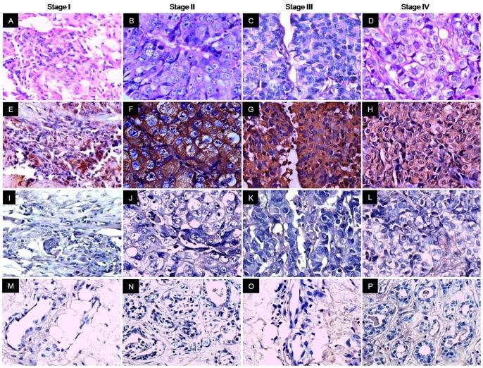
Figure 4. Immunohistochemical analysis of AKAP4 protein expression in different stages of IDC tissue specimens.
Panels A–D, H & E staining showing the histological cytostructure of representative stage I, stage II, stage III and stage IV tissue specimens respectively. Panels E–H, AKAP4 immunoreactivity was observed in the cytoplasm in all stages (brown color). Overall, 100% stage I (1/1), 86% stage II (44/51), 82% stage III (31/38) and 100% stage IV (100%) clinical specimens were found positive for AKAP4 protein expression. Panels I–L, no immunoreactivity was detected with control IgG probed specimens. Panels M- P, AKAP4 protein was not detected in any of the matched ANCT specimens probed with anti-AKAP4 antibodies. (Original magnification 400; Objective - X40).

