
Figure 3. Sucrose gradient analysis.
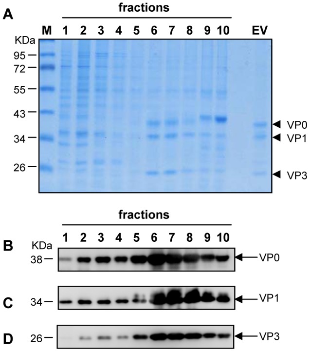
Lysates were layered onto 10–50% sucrose gradients for ultracentrifugation. Ten fractions were taken from top to bottom. (A) SDS-PAGE analysis. The ten fractions were separated by SDS-PAGE, and subsequently stained with Coomassie Blue dye. M, protein marker. EV, purified whole virion EV71 standard from Hualan Inc. (B) Western blotting with anti-VP0 polyclonal antibody. (C) Western blotting with anti-VP1 polyclonal antibody. (D) Western blotting with anti-VP3 polyclonal antibody.