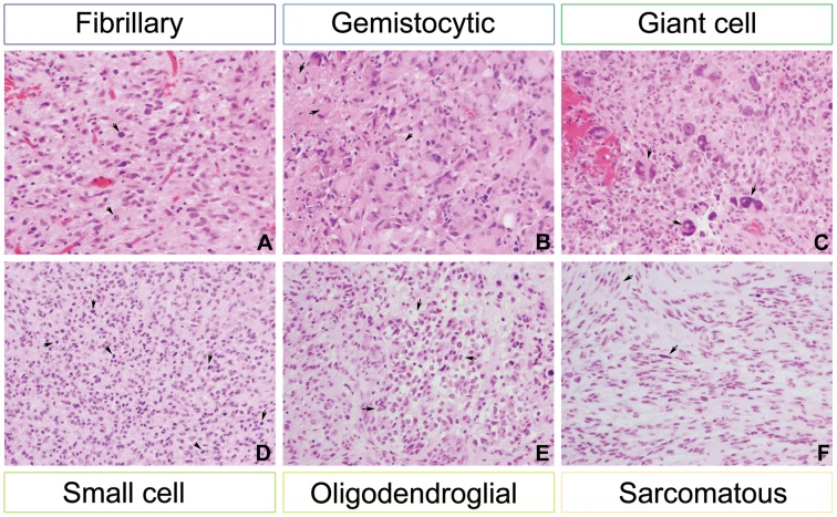
Figure 1. Distinct cell morphological types in clinical glioblastoma specimens.
Examples of tumours are shown with predominant morphology classified as fibrillary (A, RMH5690, pleomorphic cells with pink cytoplasm and mitotic figures (arrows)), gemistocytic (B, RMH6716, cells with abundant glassy cytoplasm and eccentric nuclei (arrows)), giant cell (C, RMH6004, very large pleomorphic cells with hyperchromatic nuclei (arrows)), small cell (D, RMH5723, small cells with brisk mitotic activity (arrows)), oligodendroglial (E, RMH5970, ‘fried egg’ appearance of tumour cells (arrows)), and sarcomatous (F, RMH5966, disorganised fascicles of spindle cells (arrows)). All images original magnification×200, haematoxylin and eosin.

