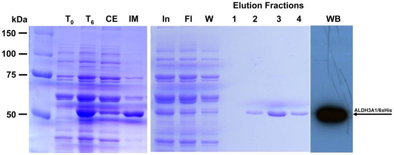
Figure 7. Protein expression and purification of recombinant his-tagged ALDH3A1.

SDS-PAGE analysis at various stages of purification of recombinant his-fused ALDH3A1 using Ni-affinity chromatography (Coomassie blue staining). T0: Total cell extract form bacterial culture co-expressing pG-KJE8 along with ALDH3A1 prior to IPTG induction, T6: Total cell extract 6 hours after IPTG induction, CE: Crude extract of lysed cells 6 hours after IPTG induction, IM: Insoluble matter of lysed cells 6 hours after IPTG induction, In: Input of the column, Fl: flowthrough part, W: wash part, Elution fractions: purified recombinant protein eluted from Ni-NTA column. WB: western immunoblotting of purified recombinant ALDH3A1/6xHis. The arrow indicates the position of the recombinant his-tagged ALDH3A1 at approximately 51 kDa.
