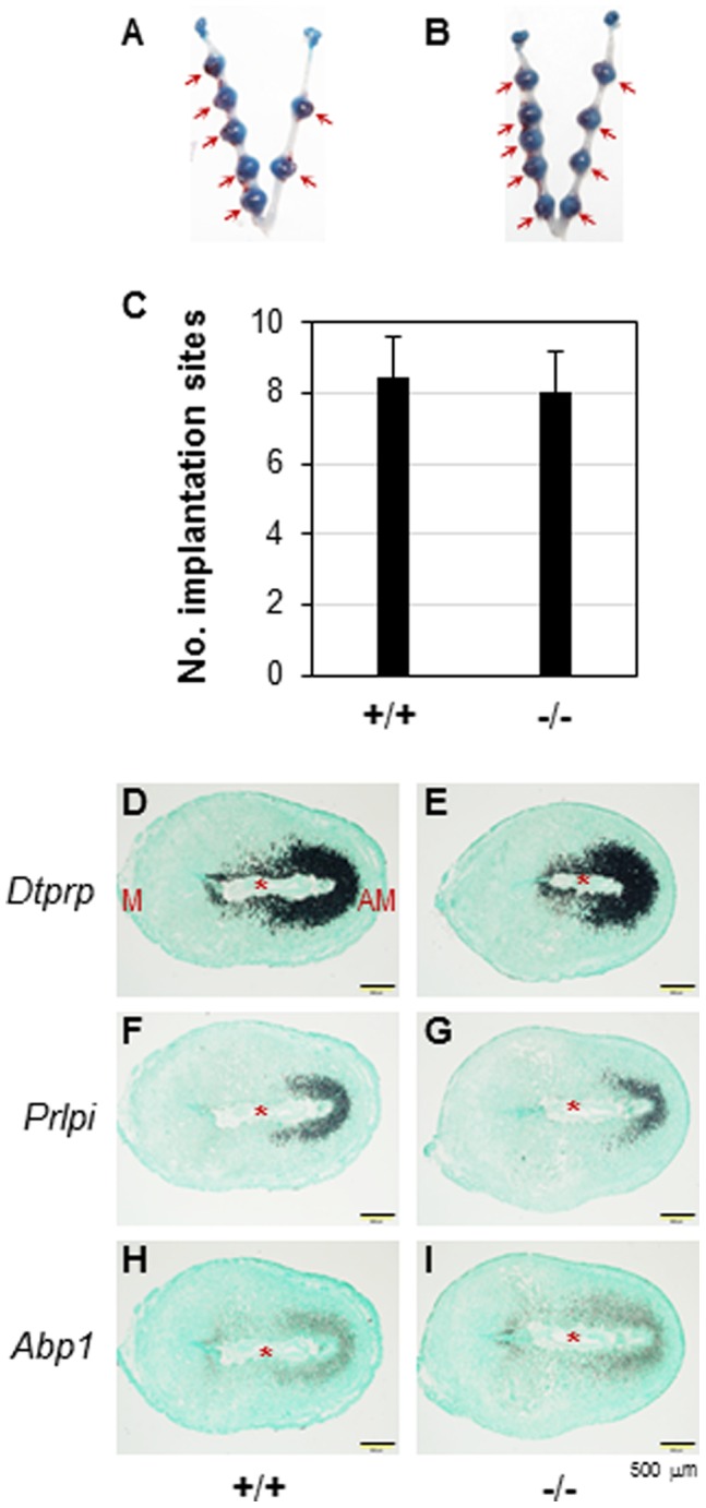
Figure 6. Deletion of Prss35 on embryo implantation and the expression of decidualization markers in D7.5 uterus.

A. A representative uterus from D7.5 WT mice. B. A representative uterus from D7.5 Prss35 (−/−) mice. A & B. Red arrows, implantation sites. C. The average number of implantation sites per mouse in the WT (+/+, N = 7) or Prss35 (−/−) (−/−, N = 7) females. All the mating males were Prss35 (−/−) for both groups except one WT male mated with one female in the WT group. Error bars, standard deviation. D∼I. Expression of decidualization markers in D7.5 uterus by in situ hybridization. D. Dtprp in WT uterus. E. Dtprp in Prss35 (−/−) uterus. F. Prlpi in WT uterus. G. Prlpi in Prss35 (−/−) uterus. H. Abp1 in WT uterus. I. Abp1 in Prss35 (−/−) uterus. Red star, embryo; M, mesometrial side; AM, antimesometrial side; scale bars, 500 µm.
