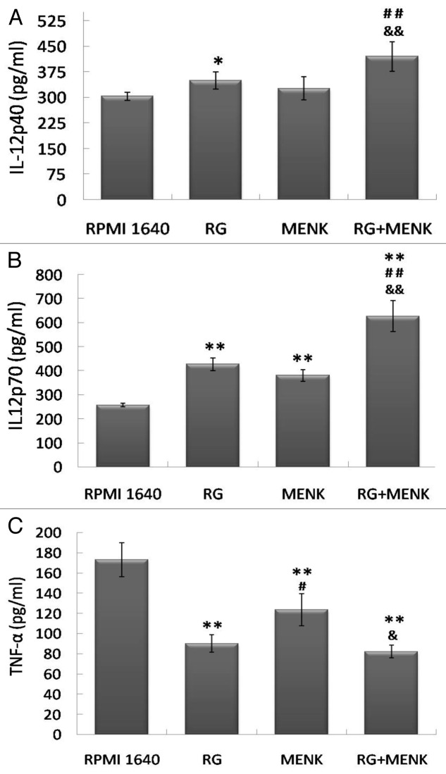
Figure 3. The production of IL-12p40 (a), IL-12p70 (b) and TNF-α (c) by BMDCs after treatment with RG and/or MENK for 48hr by ELISA. After treatment with RG and/or MENK the supernatant from cell cultures was collected and the secreted cytokines were quantified by ELISA. The histograms above showed the levels of IL-12p40, IL-12p70 and TNF-α production .Results represented the mean±SD of three independent experiments samples.

An official website of the United States government
Here's how you know
Official websites use .gov
A
.gov website belongs to an official
government organization in the United States.
Secure .gov websites use HTTPS
A lock (
) or https:// means you've safely
connected to the .gov website. Share sensitive
information only on official, secure websites.
