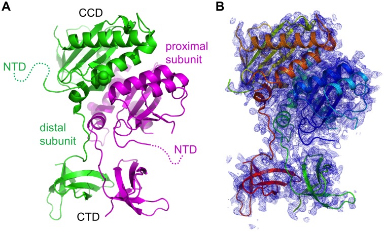
Figure 2. Overall structure of the RSV IN (1–270) dimer.
A) Ribbon diagram showing the conformation of the RSV IN (1–270)•C23S/F199K dimer in the crystal. The NTDs are poorly ordered and thus were not modeled. The positions of K199 are indicated by spheres. B) The simulated annealing composite omit 2Fo-Fc electron density map at 2.65 Å resolution, overlaid on the ribbon model. Electron density within 1.6 Å from the protein atoms is shown, contoured at 1.0σ.

