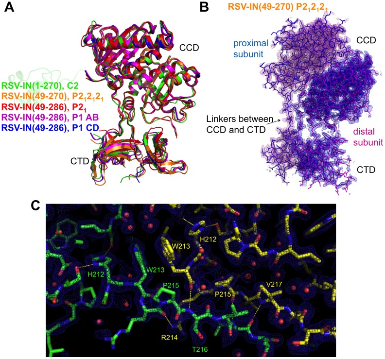
Figure 4. The asymmetric CCD-CTD dimer is a rigid entity.
A) Superposition of various RSV IN crystal structures determined in different contexts. The construct and space group for each crystal structure is indicated, with the corresponding structures color-coded. The structures of RSV IN(49–286) were reported previously [19]. The structures of RSV IN(1–270) and RSV-IN(49–270) are from the present study. An NTD in a faded color is shown to indicate that NTD is present in the crystal of RSV IN(1–270), although poorly ordered. The relative positioning of CCD and CTD is essentially the same in all crystal structures. B) The simulated annealing composite omit 2Fo-Fc electron density map calculated at 1.86 Å resolution, overlaid on the stick model of RSV IN(49–270) dimer. Electron density within 1.9 Å from the protein atoms is shown, contoured at 0.9σ. C) A close-up view of the linkers connecting CCD and CTD in the RSV IN(49–270) dimer, with the composite omit map contoured at 1.2σ. Hydrogen-bonding interactions, as described in [19], are indicated by yellow dashed lines.

