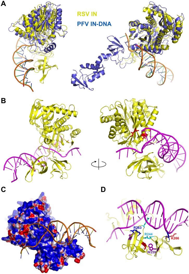
Figure 8. A hypothetical model of RSV IN-DNA complex.
A) Superposition of the CCD dimer of RSV IN onto the CCD dimer of PFV IN in the PFV IN-viral DNA-complex [7]. RSV and PFV IN proteins are colored in yellow and slate blue, respectively, and shown in two different orientations. B) PFV IN proteins were removed from the superposition in (A), leaving the bound DNA. No adjustment was made on the position or the structure of the DNA. The catalytic residues of the proximal RSV IN subunit are shown in red sticks. C) Electrostatic surface potential (positive: blue, negative: red) is displayed for RSV IN. D) The CTD residues R244, W259, R263, and K266 that have been mutated in this study, are shown in differently colored sticks.

