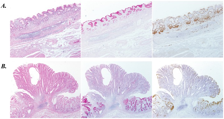
Figure 2. Immunostaining of CTSE in typical two types of gastric cancer; signet-ring cell carcinoma (A) and well-differentiated tubular adenocarcinoma (B).
HE staining (left panel), PAS staining (middle panel), and immunostaining for CTSE (right panel) were shown in sequential sections of gastric cancer specimens, where no-cancerous adjacent gastric mucosa coexist.

