
Fig. 1.
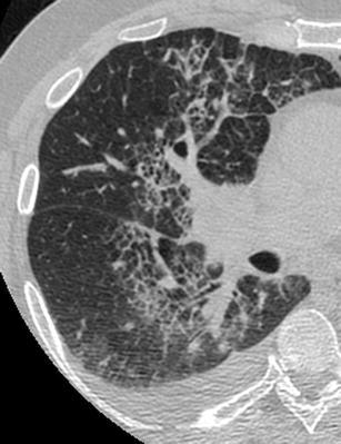
Hydrostatic pulmonary oedema secondary to endocarditis and rupture of a leaflet of the mitral valve. HRCT scan of the right lung shows a “septal pattern” characterised by thickened smoothly interlobular septae in the right parahilar area. Right pleural effusion is also seen