
Fig. 32.
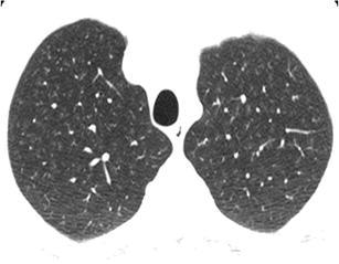
RBILD. HRCT at the level of the upper lobes exhibits an “ill-defined centrilobular nodular pattern” characterised by micronodules of ground-glass opacity that are diffusely distributed characteristically in the centre of the pulmonary lobules. In this case the history of smoking favours the diagnosis of respiratory bronchiolitis interstitial lung disease