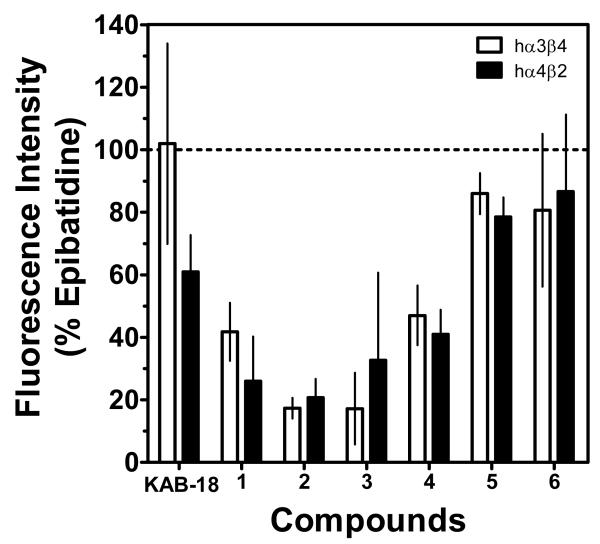
Figure 4. Effects of compounds 1–6 on recombinant nAChRs.
An inhibition assay for each compound was performed using HEK tsA201 cells stably expressing hα3β4 and hα4β2 nAChRs. Cells were loaded with Calcium 5 NW dye and stimulated with 1 μM epibatidine in the presence of a single concentration of each test compound (10 μM), as described in the Experimental Section. Results are expressed as the percentage of the control, epibatidine-stimulated peak fluorescence level. Values represent the means ± SD of three to five experiments performed in triplicate. Percentage inhibitions achieved by compounds are shown in Table 2.

