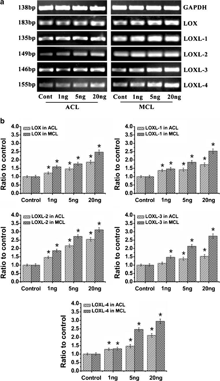
Fig. 1.
IL-1β induced dose-dependent increases of LOXs genes in both ACL and MCL fibroblasts. a Semi-quantitative PCR showed IL-1β induced higher gene expressions of LOXs in MCL than those in ACL after 2-h IL-1β treatments. Glyceraldehyde-3-phosphate dehydrogenase (GAPDH) was used as the reference gene. The gels shown were representative of four different experiments (n = 4); ACL and MCL fibroblasts in the comparison model of the experiments came from the same donors. Cont control, 1 ng, 5 ng and 20 ng 1, 5 and 20 ng/ml IL-1β, respectively. b Quantitative real-time PCR confirmed the different increases of LOXs in both ACL and MCL after 2-h IL-1β treatments. GAPDH was used as the reference gene. The △Ct method was used for measuring the fold changes. 1 ng, 5 ng and 20 ng concentrations of 1, 5 and 20 ng/ml IL-1β. The data presented were the mean of five different experiments (n = 5); scale bars SD. *p < 0.05 vs non-treated control

