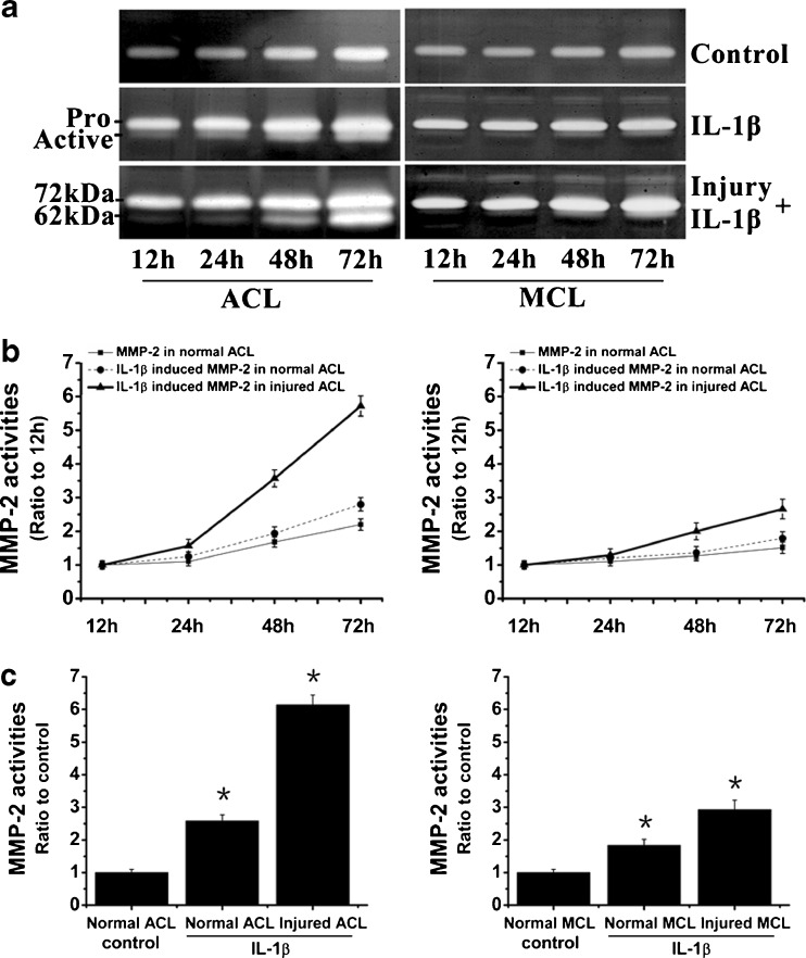
Fig. 5.
IL-1β induced higher activities of MMP-2 in injured ACL than those in injured MCL fibroblasts. a Zymography showed different expressions of MMP-2 in normal and injured ACL/MCL fibroblasts. The gels shown were representative of four different experiments (n = 4). b Quantification of MMP-2 activities showed time-dependent increases of MMP-2 activities in both normal and injured ACL/MCL. Quantification was done with Quantity One 4.6.3 software. Optical densities of the pro-MMP-2 and active-MMP-2 bands were added as the total value of activity for MMP-2. Then, the values of 24, 48 and 72 h were compared to the values of 12 h. c The indicated quantitative data refer to 72-h time points of control and treated groups, respectively. Besides, the band 62 kDa active form MMP-2 was calculated as 10 times density of the 72 kDa pro-MMP-2 band as described previously [5, 8]. The data were the mean of four different experiments (n = 4). *Significant difference with respect to control (p < 0.05)

