
Fig. 8.
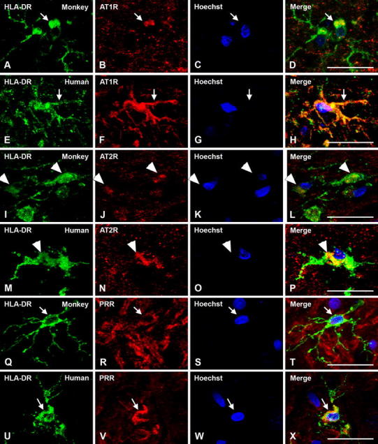
Triple immunofluorescent labeling for the microglial marker HLA-DR (green), the nuclear marker Hoechst 33342 (blue), and AT1R, AT2R or PRR receptors (red) in monkey and human substantia nigra compacta. Labeling for AT1R is apparent at the cell surface, cytoplasm and nuclear levels (a–h, arrows); labeling for AT2R is apparent mainly at the cytoplasmic level (i–p, arrowheads), and PRR mainly at the cell surface level (q–x, arrows). AT1R angiotensin II type 1 receptor, AT2R angiotensin II type 2 receptor, HLA-DR human leukocyte antigen DR, PRR prorenin/renin receptor. Scale bar 20 µm