
Fig. 2.
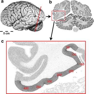
Histological procedure. a Postmortem brain sectioned in coronal plane. b Cell body-stained coronal section (20 μm) at the position marked in a. The region of interest (ROI) is labeled by the red box. c Inverted gray level index (GLI) image of the ROI with traced outer and inner cortical contours and curvilinear trajectories along the cortical ribbon. Red numbers and trajectories indicate the position along the cortical ribbon