
Fig. 4.
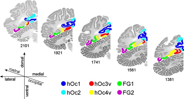
A rostro-caudal sequence of five coronal MRI sections through the left hemisphere of one single brain. Section numbers are indicated below the sections. The cortex of visual areas hOc1, hOc2, hOc3v, hOc4v, FG1 and FG2 is labeled in different colors. Distance between sections 3.6 mm