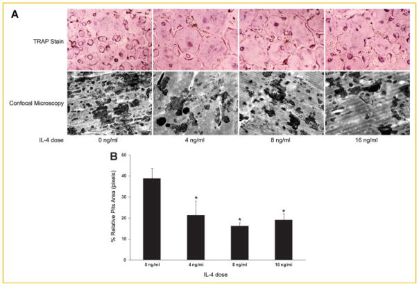
Fig. 2.
IL-4 partially impairs RANKL-induced bone resorption. A: BMMs were cultured in the presence of M-CSF and RANKL in tissue culture dishes (top panels) and on bovine bone slices (lower panels) for 36 h. The cultures were then continued in the absence and presence of IL-4 at the dose indicated. TRAP staining was performed on day 4 (top panels). Bone slices were subjected to image analysis for resorption pits as described in the Materials and Methods Section (lower panels). B: Quantification of the bone resorption assays. Four areas in the bone slices from each bone resorption assay shown in (A) were randomly chosen, and the percentage of resorbed area was determined. Data are presented as means ± standard deviation (*P <0.001 vs. absence of IL-4). No statistical significance was found between the conditions with presence of IL-4. [Color figure can be seen in the online version of this article, available at http://wileyonlinelibrary.com/journal/jcb]

