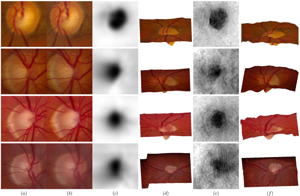
Fig. 6.
Comparison of four results obtained from the stereo fundus pairs and from the OCT scans: (a) Left fundus image centered at the optic disc, (b) right fundus image centered at the optic disc, (c) shape estimate of the optic nerve represented as grayscale maps from the OCT scans, (d) reference (left) image wrapping onto topography as output from the OCT scans, (e) shape estimate of the optic nerve represented as grayscale maps from the stereo fundus pairs, (f) reference (left) image wrapping onto topography as output from the stereo fundus pairs.

