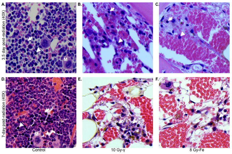
Figure 5.

Differential effects of γ and 56Fe radiation on bone marrow cells at lethal doses (n=3 mice per group, per time point, and per radiation dose). Upper panel shows bone marrow sections at 3.5 d after 10 Gy of γ (B) and 8 Gy of 56Fe (C) radiation. Lower panel shows bone marrow sections at 7 d after 10 Gy of γ (E) and 8 Gy of 56Fe (F) radiation. Panels (A) and (D) represent unirradiated control sections. Presented images are at 40X magnification. Arrow: erythroid precursor; Arrowhead: myeloid precursor; M: megakaryocyte.
