
Fig. 2.
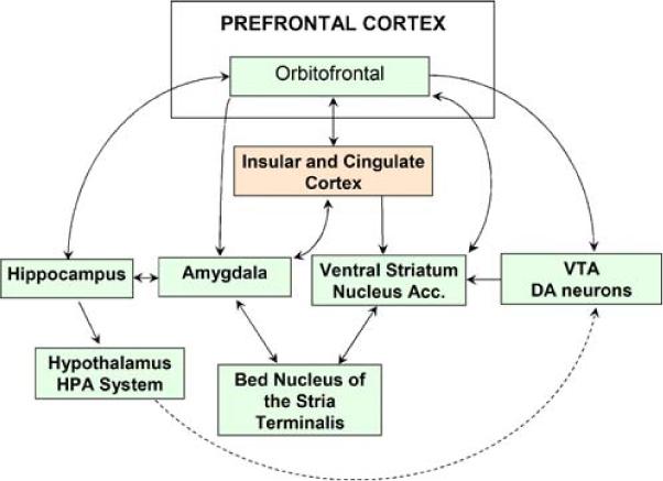
The extended limbic network consists of the basal forebrain, extended amygdala, and limbic lobe as described by Heimer and Van Hoesen (2006). To aid in visualization, not all structures or established projections are displayed. Regions of the basal forebrain, which includes areas usually referred to as the ventral striatum and nucleus accumbens, receive input from the extended amygdala, limbic lobe, and dopaminergic neurons of the ventral tegmental area. The extended amygdala includes the central and medial nuclei of the amygdala, their extensions to the bed nucleus of the stria terminalis, as well as downstream projections to the ventral striatum and nucleus accumbens. The extended amygdala has abundant associative connections with the hypothalamus as well as greater limbic lobe, including the hippocampus, cingulate and insular cortex, lateral basal cortical amygdala, and OFC