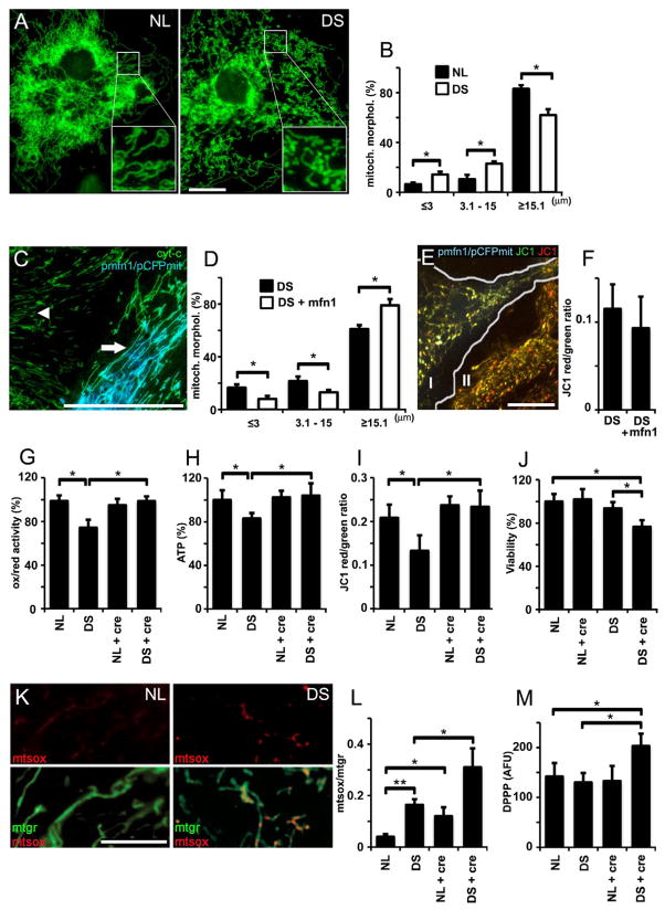
Figure 1. Mitochondrial alterations in DS astrocytes.
(A) NL and DS astrocytes were stained with Mitotracker green. Elongated mitochondria predominate in NL astrocytes (NL; inset), while the number of puncta-like and globular mitochondria was higher in DS astrocytes (DS; inset). Scale bar: 10 μm.
(B) Quantification of mitochondrial shapes binned according to length/size in NL and DS astrocytes.*p<0.05
(C) A DS astrocyte (arrow) co-expressing mitofusin 1 (pmfn1) and cyan fluorescent protein targeted to mitochondria (pCFPmit) shows tubular mitochondria. In an adjacent, non-transfected DS cell (arrowhead), cytochrome C IF (green) illustrates shorter mitochondrial shapes. Scale bar: 20 μm.
(D) Morphometric analysis shows a higher frequency of longer mitochondria and lower frequency of shorter ones in mfn1-expressing DS astrocytes. *p<0.05
(E) A DS astrocyte cotransfected with pCFPmit and pmfn1 (I) shows longer mitochondria (JC-1 green) compared to a non trasnfected astrocyte (II). The number of energized mitochondria did not change (JC-1 red). Scale bar: 10 μm.
(F) JC-1 red/green ratio shows no differences in the frequency of energized mitochondria in pmfn1-expressing astrocytes compared to non transfected cells.
(G, H and I) Quantification of ATP, ox/red activity and MMP (red/green JC-1 fluorescence ratio). Treatment with 5 mM creatine (cre) markedly increased all three parameters of mitochondrial activity in DS astrocytes.*p<0.01.
(J) Quantification of astrocyte viability (LDH). Note the reduction in viability in DS astrocytes treated with creatine (5 mM). *p<0.05
(K) NL and DS astrocytes labeled with Mitotracker green and MitoSox. Simultaneous staining allows the visualization of superoxide production (mtsox, red) in live cell mitochondria (mtgr, green). Scale bars: 20 μm.
(L) Superoxide levels expressed as a MitoSOX/Mitotracker ratio (superoxide signal/total mitochondrial signal). Superoxide in DS mitochondria was further elevated by treatment with 5 mM creatine (NL+cre, DS+cre). *p<0.05 and **p<0.01
(M) Quantification of lipid peroxidation. DPPP was used to monitor lipid peroxidation, which is expressed as arbitrary fluorescent units (AFU). DS astrocytes treated with creatine (cre, 5 mM) exhibited a significant increase in lipid peroxidation (139% ± 11.2%) *p<0.01
Statistics: (B) and (D), by Student’s t test; (G), (H), (I), (J), (L) and (N), by two way ANOVA followed by Tukey HSD test.
(G), (H), (I), (J), (L) and (M): values are the mean ± SD, n=8 independent experiments with cultures derived from different NL and DS brain specimens.

