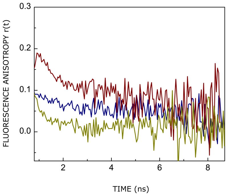
FIGURE 5.
Time-resolved fluorescence anisotropy decay of tryptophan residues in the CXCR1 N-domain peptide in AOT reverse micelles corresponding to w0 = 7 (blue) and 20 (red). The fluorescence anisotropy decay of tryptophan residues of the same peptide in bulk water is shown for comparison (olive). The excitation wavelength was 295 nm and emission was monitored at 335 and 340 nm (for reverse micellar samples corresponding to w0 = 7 and 20, respectively), with a combination of a monochromator and a 320 nm cut-off filter, using a TCSPC setup. Emission was set at 350 nm for the peptide in water. All other conditions are as in Figure 2. See Experimental Section for more details.

