
Figure 3. Dicer plays a role in modeling of the extra-cellular basement matrix of WAT.
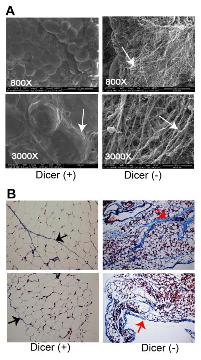
(A) Scanning electron micrograph of WAT from 12-day old Dicer-conditional mice either lacking the aP2-Cre transgene [Dicer (+)] or bearing the aP2-Cre transgene [Dicer (−)]. White arrows indicate the collagen fibers that normally form a thin basement membrane around the adipose tissue. Magnification of each sample is given. (B) Masson’s trichome staining of WAT of 12-day old Dicer-conditional mice either lacking the aP2-Cre transgene [Dicer (+)] or bearing the aP2-Cre transgene [Dicer (−)]. Collagen stains blue, the cytoplasm red, and the nuclei black. Arrows point to collagen in the basement membrane surrounding the adipocytes and in the spaces separating the depots. Magnification of Dicer (+) and Dicer (−) samples: top panels = 20X, bottom panels= 40X.