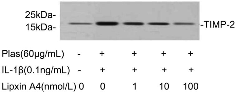
Figure 7. Effects of LXA4 on the expression of TIMP-2 by corneal fibroblasts.

Cells were cultured in the absence or presence of IL-1β (0.1ng/mL), and in the presence of the indicated concentrations of LXA4. The culture supernatants were then subjected to immunoblot analysis with antibodies to TIMP-2, Data are representative of three independent experiments. The positions of bands corresponding to the TIMP-2 are indicated on the right, and those of molecular size are shown on the left.
