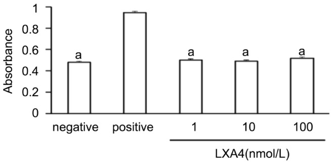
Figure 8. Lack of a cytotoxic effect of LXA4 on corneal fibroblasts.
Cells were incubated for 24 hours in MEM and in the absence (negative control) or presence of 1nmol/L, 10nmol/L or 100nmol/L LXA4, after which the culture supernatants were assayed for LDH activity with a colorimetric assay. The amount of LDH released from cells by 0.1% Triton was determined as a positive control. Data are means±SEM from three times. aP<0.05 vs 0.1% Triton (Dunnett's test).

