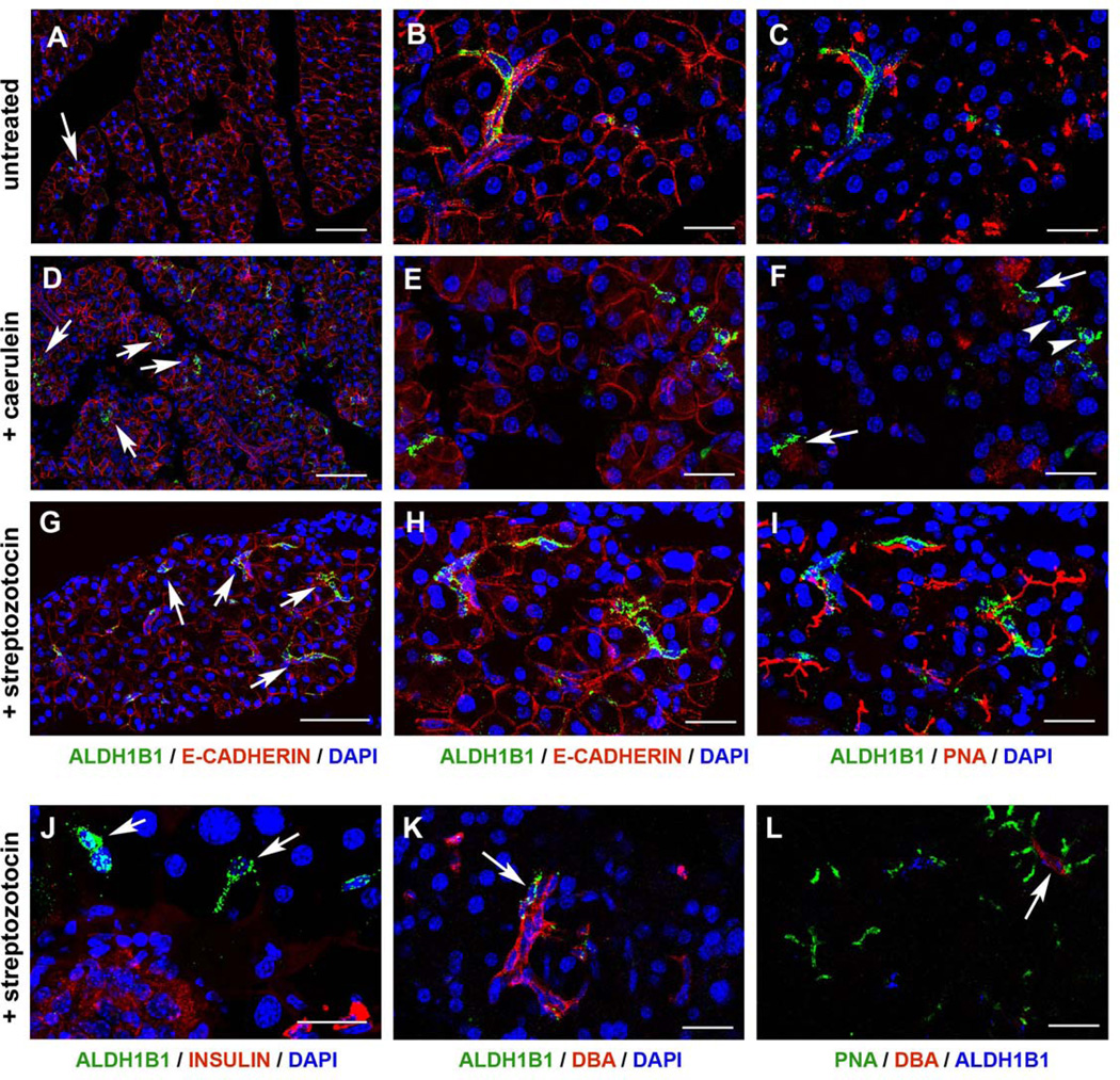
Figure 6. Very rare ALDH1B1+ cells persist in the adult pancreas but their number expands vastly following either caerulein or streptozotocin insults.
(A–C) Triple immunofluorescence of wt, untreated pancreata showed the presence of rare elongated cells expressing ALDH1B1 (A–C, arrows in A), E-CADHERIN (A, B) and PNA (C).
(D–F) The number of ALDH1B1+ cells expanded by 20 – fold in response to acute pancreatitis as shown by triple immunofluorescence for ALDH1B1 (D–F, arrows in D), E-CADHERIN (D, E) and PNA (F). Most ALDH1B1+ cells were PNA+ (F, arrows) but some were PNA− (F, arrowheads).
(G–I) The number of ALDH1B1+ cells expanded by 10 – fold in response to β cell ablation as shown by triple immunofuorescence for ALDH1B1 (G–I, arrows in G), E-CADHERIN (G, H) and PNA (I).
(J–L) ALDH1B1+ cells occasionally surrounded β cell depleted islets (J, arrows). Occasionally, ALDH1B1+/DBA+ (K, arrow) or ALDH1B1+/DBA+/PNA+ (L, arrow) cells were observed.
Scale bars: A, D, G 100 um; B–C, E–F, H–I, J–L 25 um.

