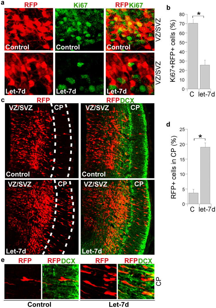
Figure 3. In utero electroporation of let-7d promotes neuronal differentiation and migration in embryonic mouse brains.
(a). In utero electroporation of let-7d into E13.5 mouse brains reduced the proliferating cell population in the ventricular zone and subventricular zone (VZ/SVZ). The Proliferating cells were labeled by Ki67. (b). Quantification of Ki67+RFP+ cells. The percentage of Ki67+RFP+ cells over total RFP+ cells in the VZ/SVZ of let-7d-electroporated brains were calculated and plotted. Error bars are standard deviation of the mean.* p<0.01 by Student's t-test. (c). Transfection of let-7d into E13.5 mouse brains induced precocious cell migration to the cortical plate. The transfected cells were shown in red due to the expression of the RFP reporter. The let-7d mutant RNA was used as the control RNA. CP stands for cortical plate; DCX stands for doublecortin. (d). Quantification of transfected cells (RFP+ cells) that migrated to the CP. C stands for the let-7d mutant control RNA. Error bars are standard deviation of the mean.* p<0.05 by Student's t-test. (e). Enlarged images of the RFP+DCX+ cells in the CP of control RNA or let-7d-transfected brains. The RFP+DCX+ cells showed yellow processes due to the merge of red and green colors.

