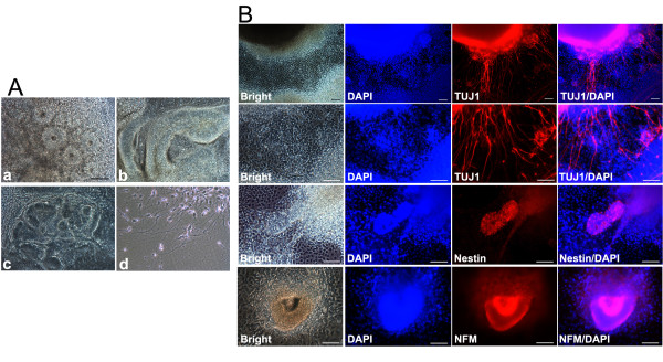
Figure 4.

EB-mediated neural differentiation of TF-SCAP iPSCs in vitro. (A-a to Ad) Four-day-old EBs were plated into Matrigel-coated wells of six-well plates. On day 8 after neurogenic stimulus, differentiated iPSCs developed into neural rosette morphology. Some cells extended elongated cell cytoplasmic processes resembling axons. (scale bar: 100 μm for all images in A) (B) Immunofluorescence analysis of differentiated neural-like cells showing positive staining of neuronal marker βIII-tubulin (TUJ1). Cells forming a neural rosette or spherical morphology were also positive for nestin and NFM. Expressed genes stained in red; DAPI, nuclear stain. (scale bar: 100 μm). DAPI, 4',6-diamidino-2-phenylindole; EB, embryoid body; iPSCs, induced pluripotent stem cells; SCAP, stem cells from apical papilla; TF, transgene free.
