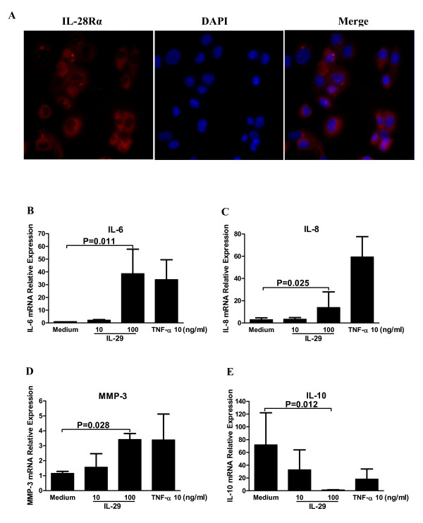
Figure 5.
IL-29 induced cytokine expression by rheumatoid arthritis (RA) synovial fibroblasts. (A) Immunofluoresence staining for IL-28Rα in MH7A cells. IL-28Rα-positive cells were stained red. The magnification was × 400. (B-E) Induction of IL-6, -8, -10 and MMP-3 in MH7A cells by IL-29. Levels of mRNA for IL-6 (B), IL-8 (C), MMP-3 (D) and IL-10 (E) in MH7A were determined by real-time PCR after 24 h incubation with IL-29 or TNF-α (a positive control). Because the effects of IL-29 at 1 ng/ml or IL-29 incubation for 48 h on the expression of the above cytokines in MH7A cells are similar to the medium control group or 24 h incubation, the data from IL-29 at 1 ng/ml or IL-29 incubation for 48 h in MH7A are not shown in the figure. IL-29 could not induce the expression of IL-17 in MH7A cells and data are not shown in the figure. Relative gene expression was determined by the 2-ΔΔct method. Values shown are the mean ± SD for five separate experiments performed in triplicate. Actual P values are shown in the graph.

