
Figure 3.
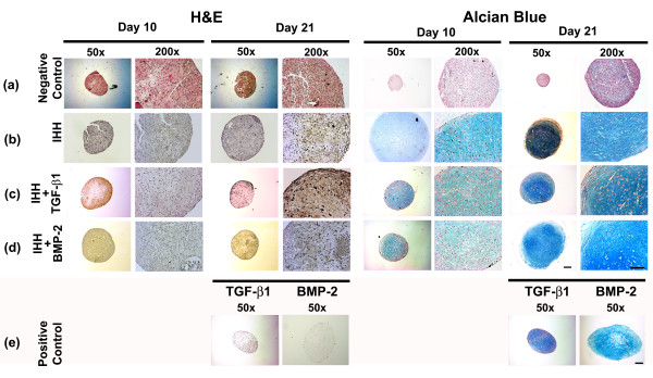
Histological assessment of chondrogenesis in mesenchymal stem cell aggregates after adenoviral Indian hedgehog gene transfer. Following genetic modification with (a) Ad.GFP (negative control), (b) Ad.IHH (IHH), (c) Ad.IHH and Ad.TGF-β1 (IHH + TGF-β1), (d) Ad.IHH and Ad.BMP-2 (IHH + BMP-2), or (e) Ad.TGF-β1 or Ad.BMP-2 only (positive control; TGF-β1 or BMP-2) at 5 × 10² virus particles/cells, mesenchymal stem cells were maintained as pellets for 3 weeks until aggregates were harvested and processed. Representative sections after 10 and 21 days are shown. Left: H & E staining for evaluation of cellularity and cell morphology. Right: Metachromatic staining with Alcian Blue for detection of matrix proteoglycans. Panels reproduced at low (50×; bar = 200 μm) and high (200×; bar = 50 μm) magnification as indicated. Ad., adenoviral vector; BMP, bone morphogenetic protein; IHH, Indian hedgehog; TGF-β, transforming growth factor beta.