Figure 6.
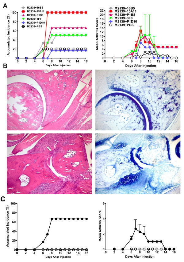
Cartilage oligomeric matrix protein (COMP)-specific mAbs mediate arthritis. (A) Accumulated incidence and severity of arthritis. Two-month-old naïve male QB F1 mice were injected intravenously (i.v.) with 9 mg of an equal combination of M2139 with a single COMP-mAb. All the mice received lipopolysaccharide (25 μg per mouse) intraperitoneally on day 5 and were scored for arthritis up to 16 days. The error bars in the severity graph indicate standard error of the mean (SEM), n = 6 per combination group. (B) Histology of tarsal joint sections. Paws of QB F1 mice on day 16 after the antibody transfer were collected, fixed, decalcified, sectioned and stained with hematoxylin/eosin (left panel) or toluidine blue (right panel). Top panel, M2139 plus PBS-injected control mice; Bottom panel, M2139 plus 15A11-injected mice. Note thickened synovial lining, inflammatory cellular infiltrate extending over the cartilage surface and erosions of cartilage and bone in animals injected with M2139 plus 15A11 (bottom). Results shown are representative of those obtained from three to four mice in each group. Infiltration cells, glycosaminoglycan loss and joint surface erosions are indicated with arrows. (C) Accumulated incidence and severity of arthritis. Two-month-old naïve male QB F1 mice were injected i.v. with 9 mg of an equal combination of 1B8, 1D10, 3F8, 15A11 and 16B5 anti-COMP monoclonal antibodies (filled circles). The same amount of a combination of isotype-matched control antibodies (L243 + G11) was injected into a separate group of animals (open circles). Results shown are pooled values from two similar experiments with balanced groups. The error bars in (A) and (B) indicate the SEM.

