
Figure 1.
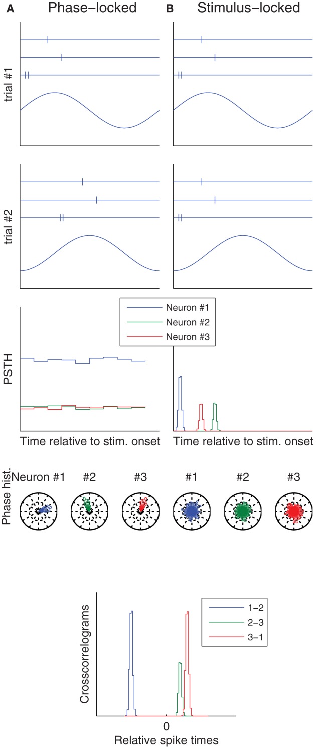
Phase vs. stimulus locking. Column (A) (resp. B) illustrates a situation in which spikes lock to an ongoing oscillation (resp. to stimulus onset). The first two rows correspond to two trials, and show both the raster plots of three neurons (top), and the ongoing oscillation (bottom), whose phase at stimulus onset is different from trial-to-trial. Post-stimulus time histograms (PSTH), which use the stimulus onset as a reference time, only reveal the temporal structure in the stimulus locked-case. Conversely, spike phase histograms, which use the oscillation peak as a reference time, only reveal the temporal structure in the phase locked-case. Spike time cross-correlograms between pairs of neurons reveal the temporal structure in both cases. These are good news, because downstream neurons only care about relative spike times—they ignore both the stimulus onset time and the oscillation phase.