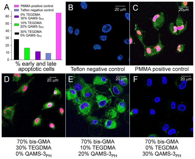
Figure 7.
A. Distribution of early and late apoptotic MDPC-23 cells, after exposure to i) Teflon negative control; ii) Polymethyl methacrylate (PMMA) positive control; iii) Polymerized resin composed of bis-GMA:TEGDMA:QAMS-3PH with a mass ratio of 70:30:0; iv) Polymerized resin composed of bis-GMA:TEGDMA:QAMS-3PH with a mass ratio of 70:10:20; and v) Polymerized resin composed of bis-GMA:TEGDMA:QAMS-3PH with a mass ratio of 70:0:30. The presence of later apoptotic cells even in the negative control group may be attributed to membrane damage to some of the cells (hence, stainable with Annexin-V) when trypsin was employed for cell detachment from the culture plates. Despite this limitation, comparison of the percentage of early and late apoptotic cells shows that the 70:0:30 polymerized resin group has the lowest % of apoptotic cells among the three resin groups. B-F. Two-photon laser fluorescence microscopy imaging of MDPC-23 cells after exposure to eluents derived from 5 groups. The nuclei of healthy cells were stained positively with Hoescht 33342 (stains DNA of both vital and non-vital cells; blue fluorescence) only. The nuclei of dead cells were stained also positively with ethidium homodimer-III (Etd-III, non-vital DNA marker; red fluorescence). Merging of the channels results in pink nuclei due to combination of the blue and red fluorescence. The cytoplasm of apoptotoic cells were stained positively with FITC-Annexin V (stains cytoplasmic phosphatidylserine; green fluorescence). B. Only healthy cells (stained by Hoechst only, not by FITC-Annexin V and EtD-III) are seen in the Teflon negative control (absence of apoptosis or necrosis). C. Cells were stained blue, green and red in the PMMA positive control, which is indicative of late apoptotic cells or dead cells progressing from the apoptotic cell populations. D. Similar dead cells were seen in the experimental resin group with bis-GMA/TEGDMA/QAMS-3PH mass ratio of 70:30:0. E. Early apoptotic cells (stained both blue and green) observed in the in the experimental resin group with bis-GMA/TEGDMA/QAMS-3PH mass ratio of 70:10:20. F. Most of the cells are healthy (stained blue only) in the experimental resin group with bis-GMA/TEGDMA/QAMS-3PH mass ratio of 70:0:30. Some early apoptotic cells are also present.

