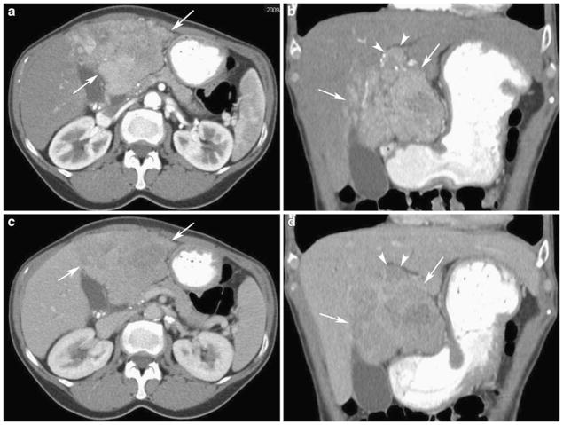
Fig. 1.
Fifty-five-year-old woman with hepatitis C, cirrhosis, and hepatocellular carcinoma. Axial (a) and coronal (b) contrast-enhanced CT in the arterial phase shows a heterogeneously enhancing liver mass in the left hepatic lobe with ill-defined margins (arrows). There is enhancing tumor thrombus in the portal vein (arrowheads). Axial (c) and coronal (d) contrast-enhanced CT in the portal venous phase shows an infiltrative mass in the left hepatic lobe (arrows) with areas of washout consistent with hepatocellular carcinoma

