
Fig. 3.
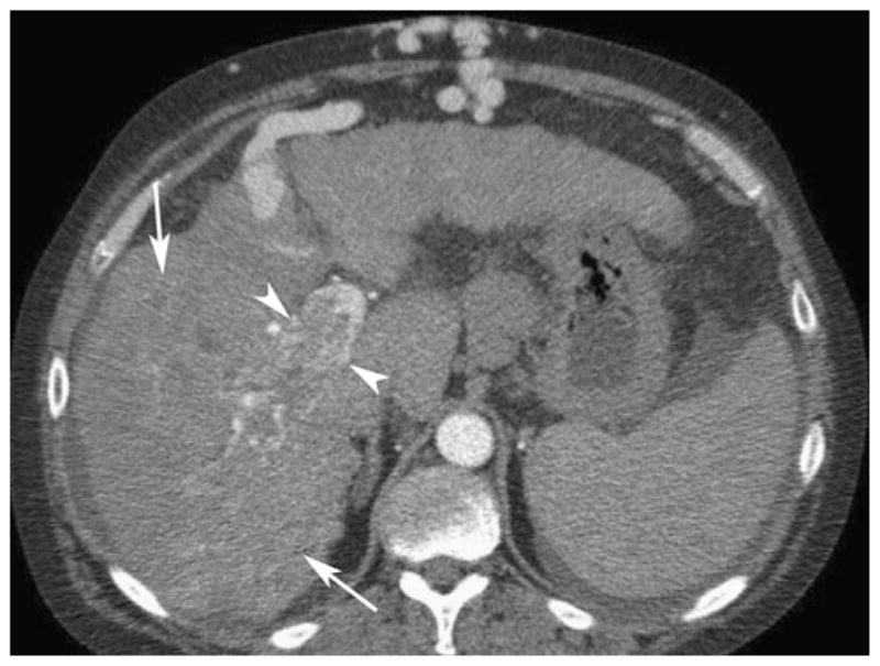
Fifty-three-year-old man with alcoholic cirrhosis and hepatocellular carcinoma. Axial contrast-enhanced CT in the arterial phase shows thrombus in the right portal vein (arrowheads). Small enhancing vessels seen within the thrombus indicate the presence of tumor. The infiltrative HCC (arrows) in the right hepatic lobe is difficult to appreciate. Stigmata of portal hypertension are seen, including ascites and caput medusa