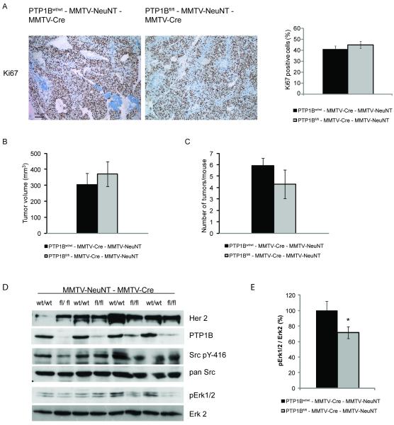
Figure 2. Epithelial deletion of PTP1B does not affect proliferation and signaling of NeuNT-induced mammary tumors.
A. Immunohistochemical analysis of mammary tumors from PTP1Bwt/wt - MMTV-Cre - MMTV-NeuNT and PTP1Bfl/fl - MMTV-Cre - MMTV-NeuNT mice 5 weeks after their onset. Representative images of Ki67-stained sections of mammary tumors as indicated. Bar graph showing the quantification of Ki67 staining in mammary tumors (n=6). P=0.41 student’s t-test.
B, C. Tumor volume and the number of tumors per mouse of PTP1Bwt/wt - MMTV-Cre - MMTV-NeuNT (n=12) and PTP1Bfl/fl - MMTV-Cre - MMTV-NeuNT mice (n=6) 30-35 days after tumor onset. P=0.56 for tumor volume and P=0.24 for the number of tumors per mouse, student’s t-test.
D. Immunoblots of mammary tumor lysates from PTP1Bwt/wt - MMTV-Cre - MMTV-NeuNT and PTP1Bfl/fl - MMTV-Cre - MMTV-NeuNT mice 5 weeks after tumor onset.
E. Densitometric quantification of pErk1/2 normalized to Erk2 levels (n=4). P=0.03, student’s t-test.

