
Figure 2.
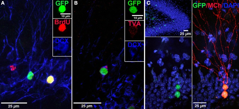
Labeling of “starter” neural progenitor cells. (A) Photomicrograph showing retrovirally labeled newborn GCs (pRV-SYN-GTRgp) expressing nucleus-localized histone-tagged green fluorescent protein (hGFP), avian TVA receptor, and rabies glycoprotein under control of the synapsin promoter. Co-labeling of hGFP+ cells (green), with bromodeoxyuridine (BrdU, red), and the immature neuronal marker doublecortin (DCX, blue). Insert shows a new GC expressing all three markers (yellow). (B) Confocal image of hGFP+ cell (green) co-labeled with DCX (blue) and the TVA receptor (red). (C) Overview of the DG showing newborn GCs labeled with retrovirus expressing nuclear hGFP (green, top). Magnification of the GC layer shows dual virus labeled newborn “starter” cells with retrovirus expressing hGFP (green) and EnvA pseudotyped rabies virus expressing MCherry (MCh, red) at 30 dpi. Scale bar, 25 μm. Nuclei labeled with DAPI (blue). From Vivar et al. (2012); Reprinted with permission from Macmillan Publishers Ltd.: Nature Communications Copyright 2012.