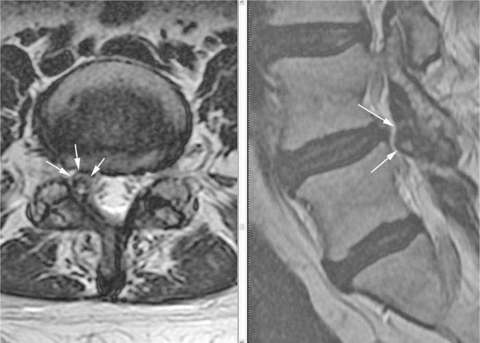
Figure 1.
Axial and parasagittal FRFSE (fast relaxation fast spin echo) T2 sequences show a cystic structure with hypointense rim and hyperintense content projecting ventrally from the right L4–5 facet joint and into the right lateral recess (white arrows). There is a mild disc bulge at this level (not visualized in this image).

